Overview
PET-CT is a nuclear medicine method that enables the physiological processes of the body to be monitored in 3D and has been used for years. In 2000, it was selected as “invention of the year” in the field of medicine by TIME magazine and broke new ground in the field of medical imaging.
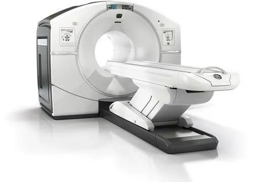
PET-CT, which consists of combining PET device with CT, provides anatomical and physiological information in a single session and increases the accuracy of the examination.
The usage area of PET-CT is generally in the field of oncology. It is used in diagnosis, staging, restaging, and evaluation of response to treatment of lymphoma, malignant melanoma, head and neck tumors, lung cancers, breast cancer, colorectal cancers, esophagus, and stomach tumors, gynecological cancers, mesothelioma, primary bone tumors, and metastases of unknown primary origin.
PET-CT scan also yields results in neurological cases such as early diagnosis of Alzheimer’s disease, determination of epilepsy focus, and the investigation of the presence of living tissue in the heart after a heart attack.
The FDG, which is required for the PET-CT examination, is administered intravenously and the accumulation in cells with high metabolic activity such as tumor cells results in imaging successfully.
Since the image is taken from the whole body, even cases such as the spread of cancer to another part of the body (metastasis) can be detected.
Today, early diagnosis of many diseases is easier with PET-CT. In the pre-PET-CT period, whether the nodules in the body are cancerous or not can only be determined by biopsy, now PET-CT can determine whether the lesions are cancer or not with high sensitivity.